


In order to see the pathological changes in the condition of the nails and the skin of the feet in time and start treatment as soon as possible, it is important to know what the fungus of the nail plate looks like. The faster steps are taken to eliminate the disease, the more likely it is that it will be able to prevent the destruction of the nail plate and restore its normal appearance. Find out how the fungus manifests itself at different stages and what are the characteristics of the course of this disease.
What does onychomycosis look like?
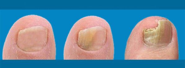
To understand that the nail plate has been infected with a fungal infection (onychomycosis), you need to know what a healthy nail looks like. In normal conditions, the nail is smooth, the horny plate is pale pink, smooth, without dents, bumps or delamination. Healthy nails are strong and elastic, not thickened. But changes in their appearance can indicate many pathological processes in the body, so it is necessary to identify the specific symptoms that exist in onychomycosis. They may vary depending on the form of the disease.
- Normotropic.This is the initial stage of nail fungus. Horny plates change color, yellowish and white spots and stripes appear on them, as well as an unpleasant smell. This is the initial stage of the disease, so the nails maintain a normal thickness and a relatively healthy appearance. This stage begins to appear at the end of the incubation period.
- Hypertrophic: the color changes more, the plate begins to thicken, and the luster disappears. Changes in shape and partial destruction of the plate along the edges can be observed.
- Atrophic:The affected nail is separated from the nail bed.
Another classification also depends on the appearance of nail fungus. It involves dividing the infection into several types depending on the part of the nail affected by the fungus:
- Distal.There is delamination and yellowing of the edge of the plate, keratinization of the nail bed. In some cases, the nail may be completely affected, and the root (matrix) may also be infected. Plate thinning may occur.
- Surface.The fungus affects the upper part of the horny plate, causing the appearance of white stripes and spots that turn yellow and grow over time. They can be easily removed by scraping. The plate has a loose structure. This variety is specific: this is how toenail fungus manifests itself.
- proximal.Fungus occurs under the nail, causing damage to the matrix and tissue around the plate. Cuticle rejection may occur. Deep grooves and irregularities appear on the nails.
- Amount.Nails acquire a gray-yellow color, become very thick and peel off. The plate is completely or partially destroyed.
Foot fungus
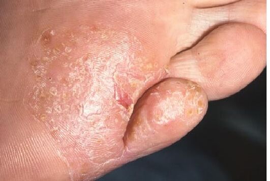
Often, the fungus on the toenail spreads to the skin of the foot. What does foot fungus look like?
In the first stage, the infection manifests itself in the form of redness and swelling of the skin, and the appearance of small cracks.
Often, changes can be seen between the toes and on the heel.
The next symptom of foot mycosis is the appearance of spots on the skin, which soon begin to itch and peel. Over time, the size of these spots increases, involving larger areas of the skin in the fungal process. There is an unpleasant smell from the feet, even if you are not wearing shoes. If treated incorrectly or untimely, foot fungus can develop into an extensive form, where deep cracks form at the base of the toes and between them, on the arch of the foot and on the heel. In addition, this stage is characterized by severe skin separation.
Diagnosis of fungal nail infection
Any person who is far from medicine can suspect a nail or foot fungal infection if he has at least a weak understanding of this disease. However, only a qualified specialist can make an accurate diagnosis and prescribe the appropriate treatment based on external examination, patient surveys, and data from the study of affected nails under a microscope. In this case, you should contact a dermatologist.
To determine whether the patient really has a fungal infection, a scraping is taken from the affected nail in the laboratory and, after placing the material in an alkaline environment, it is examined for the presence of fungal mycelium under a microscope. If certain structures are found, the diagnosis will be fully confirmed. Additional studies may be prescribed to determine the specific type of fungus; this is necessary to choose the most effective drug against the infection.
Nail fungus not only spoils the appearance of the hands and feet, but can also lead to unpleasant consequences, including the complete loss of the nail plate and the penetration of fungal infections into the body. In addition, onychomycosis and foot fungus are infectious diseases, so at the first symptoms you need to consult a doctor as soon as possible to protect your loved ones. The incubation period of the fungus can take several weeks, so the disease does not appear immediately. The sooner you get help from a specialist and accurately diagnose the disease, the faster the treatment will take place and the less money you will have to spend on expensive drugs with antifungal action.